Introduction
Medical imaging plays a crucial role in diagnosing and monitoring diseases, but it also exposes patients to ionizing radiation. While the benefits of imaging far outweigh the risks in most cases, it is essential to minimize radiation exposure without compromising diagnostic quality. One of the key concepts in optimizing radiation dose is the use of Dose Reference Levels (DRLs).
What Are Dose Reference Levels (DRLs)?
Dose Reference Levels (DRLs) are established dose thresholds that serve as a guideline for optimizing radiation exposure in medical imaging. They are not strict regulatory limits but rather reference points to help identify unusually high or low radiation doses during standard imaging procedures.
DRLs are typically set based on the 75th percentile of the dose distribution observed in clinical practice for a specific examination, ensuring that most procedures remain within a safe and reasonable range.
Importance of DRLs
- Patient Safety: DRLs help prevent unnecessary radiation exposure, reducing the potential risks associated with ionizing radiation, such as radiation-induced cancers.
- Quality Assurance: By monitoring doses, radiology departments can ensure imaging quality remains optimal while keeping doses as low as reasonably achievable (ALARA principle).
- Standardization: DRLs provide a benchmark for comparing doses across different institutions, helping identify areas for improvement.
- Regulatory Compliance: Many national and international health organizations recommend using DRLs to ensure safe imaging practices.
Establishing DRLs
DRLs are determined through a systematic process involving:
- Data Collection: Gathering radiation dose data from various healthcare facilities for specific imaging procedures.
- Statistical Analysis: Determining the 75th percentile of the dose distribution.
- Review and Validation: Experts analyze the data to ensure the reference levels are appropriate and achievable.
- Implementation and Monitoring: Facilities compare their dose levels to DRLs and adjust protocols if necessary.
DRLs are often categorized based on patient groups (e.g., adults, children) and imaging modalities, including:
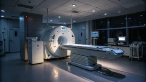
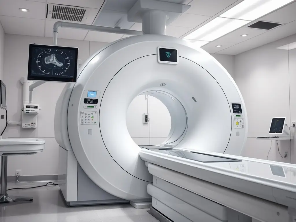
- Computed Tomography (CT): CTDIvol (Computed Tomography Dose Index) and DLP (Dose Length Product)
- Fluoroscopy: Dose-Area Product (DAP)
- Radiography: Entrance Surface Dose (ESD)
- Mammography: Mean Glandular Dose (MGD)
International Guidelines on DRLs
Organizations such as the International Commission on Radiological Protection (ICRP), International Atomic Energy Agency (IAEA), and World Health Organization (WHO) provide recommendations for setting and updating DRLs. Countries may adopt or modify these recommendations based on their healthcare systems and technological advancements.
Implementing DRLs in Clinical Practice
To effectively use DRLs, healthcare facilities should:
- Regularly monitor radiation doses and compare them with established DRLs.
- Optimize imaging protocols to minimize dose without compromising diagnostic accuracy.
- Train radiology staff on dose optimization strategies.
- Use dose-tracking software for real-time dose management.
- Conduct periodic audits to identify deviations and implement corrective actions.
Conclusion
Dose Reference Levels (DRLs) are vital tools in radiology imaging, promoting patient safety and dose optimization. By following DRLs, healthcare providers can ensure that radiation exposure remains within acceptable limits while maintaining high-quality imaging standards. Regular review and adherence to DRLs contribute to better patient outcomes and enhanced radiological practices globally.
For more information on setting up DRL contact us: info@oncorace.com